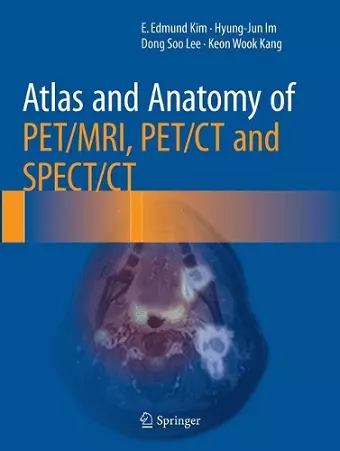
Atlas and Anatomy of PET/MRI, PET/CT and SPECT/CT
A comprehensive resource for hybrid imaging interpretation.
E Edmund Kim author Dong Soo Lee author Hyung-jun Im author Keon Wook Kang author
Format:Paperback
Publisher:Springer International Publishing AG
Published:12th Jun '18
Currently unavailable, and unfortunately no date known when it will be back

This comprehensive atlas aids in the interpretation of hybrid imaging techniques, enhancing understanding for professionals in various medical fields, including those utilizing Atlas and Anatomy of PET/MRI, PET/CT and SPECT/CT.
This atlas serves as an essential guide to understanding cross-sectional anatomy, specifically tailored for interpreting images produced by PET/MRI, PET/CT, and SPECT/CT technologies. These advanced hybrid imaging modalities are at the cutting edge of nuclear and molecular imaging, allowing for enhanced data acquisition that significantly aids in both diagnosis and treatment. By enabling the simultaneous assessment of anatomical and metabolic information, this resource addresses complex clinical scenarios and bolsters the confidence of medical professionals when interpreting scans.
The atlas is richly illustrated with high-resolution images from PET/MRI, PET/CT, and SPECT/CT, offering precise morphologic details applicable to the entire body as well as targeted regions such as the head and neck, abdomen, and musculoskeletal system. This comprehensive visual reference is designed to meet the needs of a diverse audience, including physicians and residents specializing in nuclear medicine, radiology, oncology, neurology, and cardiology.
Atlas and Anatomy of PET/MRI, PET/CT and SPECT/CT stands out as a unique and invaluable resource, bridging the gap between complex imaging techniques and practical clinical application. Whether for educational purposes or practical reference, this atlas is an indispensable tool for those involved in the interpretation of hybrid imaging studies.
“The book should be especially helpful to nuclear medicine and radiology residents, who may use it as a great source of ready-to-use knowledge in everyday clinical practice. It may also be useful to both undergraduate and postgraduate students, who will appreciate its importance as a good resource to improve their skill in interpreting hybrid nuclear medicine images. … very valuable in all imaging departments with hybrid machines in helping to improve the interpretation of individual cases in routine practice.” (Giuseppe Danilo Di Stasio and Luigi Mansi, European Journal of Nuclear Medicine and Molecular Imaging, Vol. 45, 2018)
ISBN: 9783319803999
Dimensions: unknown
Weight: unknown
594 pages
Softcover reprint of the original 1st ed. 2016