Weir & Abrahams' Imaging Atlas of Human Anatomy
Marios Loukas author Lonie R Salkowski author Tom Turmezei author Jamie Weir author Peter H Abrahams author Jonathan Spratt author
Format:Paperback
Publisher:Elsevier Health Sciences
Published:30th Jun '20
Should be back in stock very soon
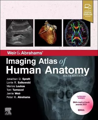
BMA Book Awards - Winner of Basic and Clinical Sciences category! The perfect up-to-date imaging guide for a complete and 3-dimensional understanding of applied human anatomy Imaging is ever more integral to anatomy education and throughout modern medicine. Building on the success of previous editions, this fully revised sixth edition provides a superb foundation for understanding applied human anatomy, offering a complete view of the structures and relationships within the whole body, using the very latest imaging techniques. All relevant imaging modalities are included, from plain radiographs to more advanced imaging of ultrasound, CT, MRI, functional imaging and angiography. Coverage is further enhanced by a carefully selected range of BONUS electronic content, including clinical photos and cases, ultrasound videos, labelled radiograph 'slidelines', cross-sectional imaging stacks and test-yourself materials. Uniquely, key syllabus image sets are now highlighted throughout to aid efficient study, as well as the most common, clinically important anatomical variants that you should be aware of. This superb package is ideally suited to the needs of medical students, as well as radiologists, radiographers and surgeons in training. It will also prove invaluable to the range of other students and professionals who require a clear, accurate, view of anatomy in current practice. Fully revised legends and labels and new high-quality images-featuring the latest imaging techniques and modalities as seen in clinical practice Covers the full variety of relevant modern imaging-including cross-sectional views in CT and MRI, angiography, ultrasound, fetal anatomy, plain film anatomy, nuclear medicine imaging and more - with better resolution to ensure the clearest anatomical views Core syllabus image sets now highlighted throughout-to help you focus on the most essential areas to excel on your course and in examinations Unique summaries of the most common, clinically important anatomical variants for each body region-reflects the fact that around 20% of human bodies have at least one clinically significant variant New orientation drawings-to help you understand the different views and the 3D anatomy of 2D images, as well as the conventions between cross-sectional modalities Ideal as a stand-alone resource or in conjunction with Abrahams' and McMinn's Clinical Atlas of Human Anatomy-where new links help put imaging in the context of the dissection room Now a more complete learning package than ever before, with superb BONUS...
"The textbook and electronic resources complement each other and enhance the learning experience of users." -© Doody's Review Service, 2021, Brian R. MacPherson, PhD (University of Kentucky College of Medicine) Score: 92-4 Stars!
ISBN: 9780702079269
Dimensions: unknown
Weight: 1020g
288 pages
6th edition